Contact: Mark Surman
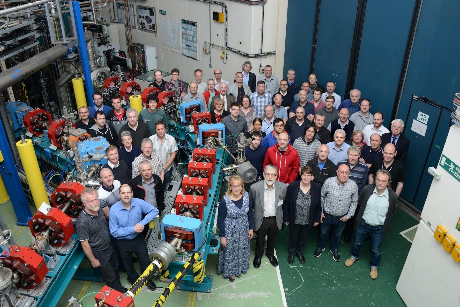
The ALICE Accelerator at Daresbury Laboratory
ASTeC successfully commissioned and operated the Energy Recovery Linac ALICE which provides three light sources:
(1) Coherent Synchrotron radiation long wavelength THz
(2) Free Electron Laser (FEL) mid-infrared and
(3) Compton generated X-rays.
Whilst the initial programmes were predominantly in accelerator and Free Electron Laser (FEL) physics, photon science exploitation was a key component from the start.
In recent years, Peter Weightman from the University of Liverpool has led a multidisciplinary team including clinicians in using the Free Electron Laser to chemically map tissue samples to investigate various cancers. This community demanded extreme high stability in both the wavelength and intensity output of the FEL. This challenge being met by MARS FEL scientists together with colleagues in other ASTeC groups.
MARS group scientists ensured world leading FEL stability for imaging experiments on ALICE, and, together with colleagues in other ASTeC groups, ran ALICE as a facility.
UK Experiments at Overseas XFELs
Immediately the world’s first X-ray Free Electron Laser, LCLS at Stanford, began user operations it produced new science and it continues to produce new science. Other facilities followed, the latest being the European XFEL. This, the world’s first superconducting XFEL, opened to users in summer 2017. As with ALICE, it is the user experience and demand for ever better performance that is driving the designs for the next generation of XFELs. MARS is involved in a number of user programmes at these XFELS. David Holland (MARS honorary scientist) works at LCLS to probe gas phase molecules undergoing UV induced fragmentation. MARS funds PhD studentships in structural biology with Prof. Samar Hasnain (University of Liverpool) working at SACLA (Japan) and Prof. Jon Marangos (Imperial College) working at at LCLS and SACLA. The Marangos work has demonstrated the huge potential for machine learning to predict XFEL pulse properties [Sanchez-Gonzalez et al., Nature Communications, 8 (2017) 15461].
The VELA Accelerator at Daresbury - capturing structure on ultrafast timescale
Knowledge of the structure of a material is key to understanding its properties and knowledge of how this structure changes is key to understanding its chemistry. If the structure can be determined on a very fast time scale (sub 100 fs) then it becomes possible to follow molecular structure evolution on the timescale of the making and breaking of chemical bonds. X-ray FELS such as the European facility in Hamburg allows such measurements.
Electrons also diffract by material and such electron diffraction has some distinct advantages over X-ray diffraction. Electron scattering cross-sections are up to a million times larger than X-rays so smaller crystals are required. Electrons also deposit much less energy into the sample to cause damage.
We have obtained diffraction from simple materials on VELA with single low charge electron bunches. These bunches have a duration of sub-100 fs at the gun but somewhat longer at the current sample position used for proof of principle experiments.
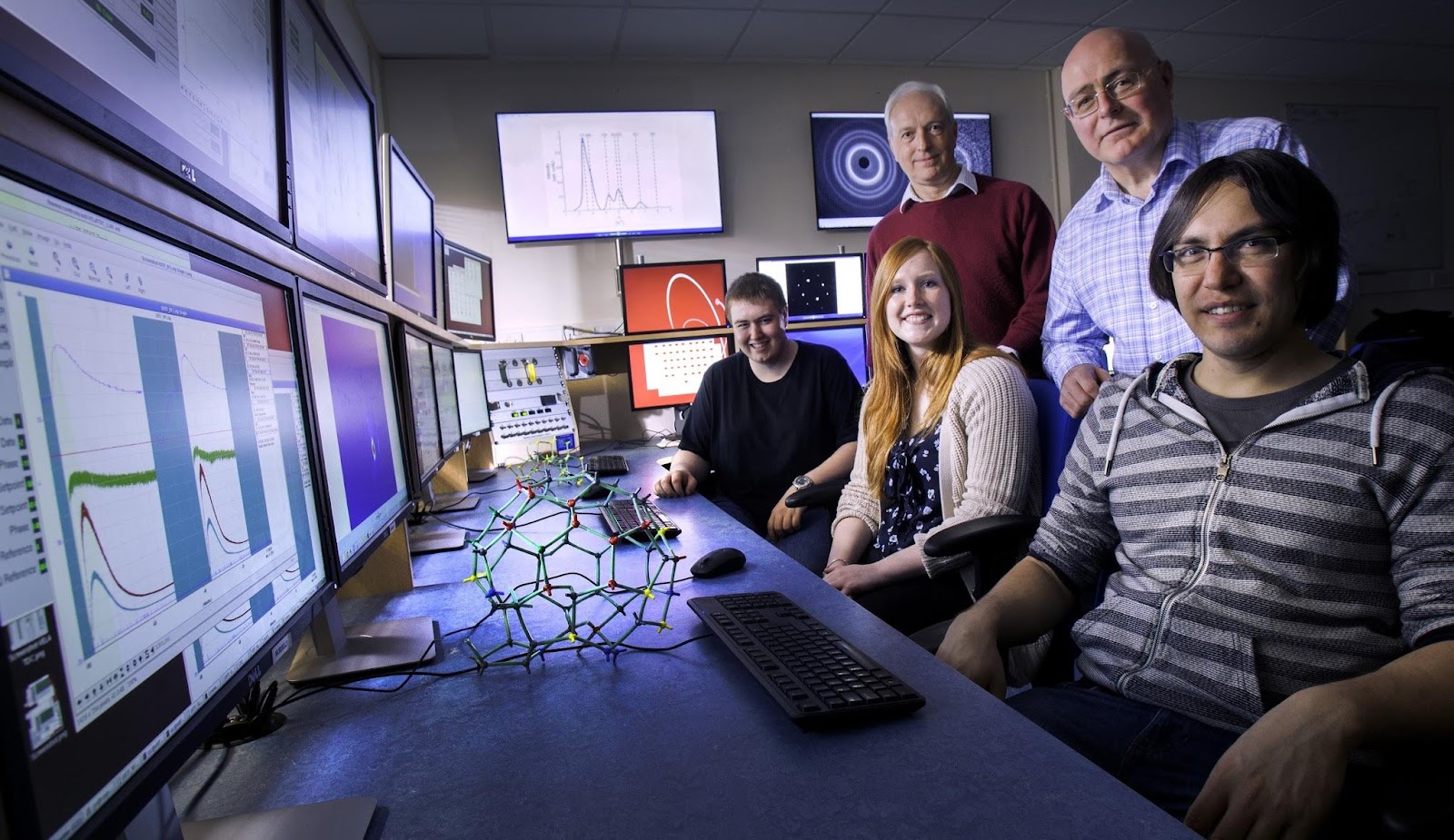
MARS and AP scientists including year in industry students in the VELA control room running Ultrafast Electron Diffraction
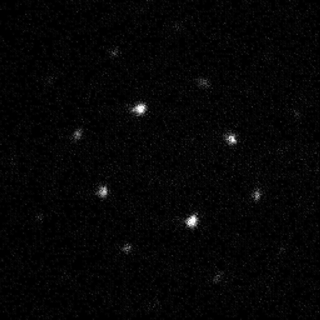
Diffraction pattern from single crystal gold obtained with a single 40 fC electron bunch with a duration ~ 100 fs